Trending

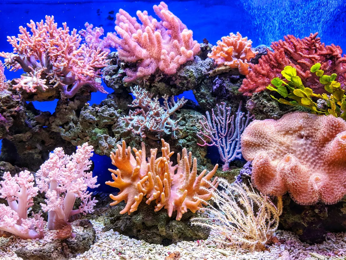
Few things irritate a reef keeper quite like a frag plug with a label faded into a grey smear. The coral is healthy, growing, and completely anonymous. Identification is not about memorising hundreds of species names. It is about narrowing things down: stony or soft, big polyp or small, branching or plating — and then checking the fine detail that separates two similar-looking genera.
Almost every coral you will meet in a home aquarium falls into one of three practical groups: soft corals, LPS (large polyp stony) and SPS (small polyp stony). These are hobby shorthand rather than strict science, but they are a genuinely useful first sorting step.
Before you think about colour, think about the body of the coral.
A quick test: does it hold its shape when lifted clear of the water? If it collapses into a wet rag, you have a soft coral. If it keeps its form and feels like rock under a thin skin, it is stony.
Step back and look at the colony's outline, especially the growing edge.
Branching forms throw up fingers or antler-like stems. Acropora is easy to spot here because each branch ends in a pale axial corallite where new growth happens; Stylophora and some Montipora branch too but tend to look blunter and more clustered. Plating forms spread outward in horizontal shelves, with Montipora capricornis as the classic example. Encrusting forms hug the rock and follow its contours. Massive forms grow in slow, rounded lumps, as Porites and many Favia do. Meandering or brain-like forms have corallites fused into winding valleys — Platygyra, Merulina and Leptoria all show this pattern. Solitary corals such as Fungia and Trachyphyllia live as a single large polyp that can rest free on the sand.
Soft corals have their own recognisable shapes. Sarcophyton and Lobophytum form a stem topped with a broad cap. Sinularia grows as branching fingers or lobes. Xenia pulses on a stalk. Clavularia and Briareum creep as mats across rock.
Take three shots: top down, side on, and a close-up with the polyps retracted. Resting a ruler or a coin beside the coral gives you scale, which matters far more than it sounds when you are comparing a frag to a photograph weeks later.
Retraction is a gift. With the polyps pulled in, you can see what the coral is actually built from.
Look at the corallites. How large are they? How close together? Do they share walls, or does each sit in its own cup with a groove between? Traditionally, Favia corallites have separate walls with a visible gap, while Favites corallites share a common wall. That single difference has saved many hobbyists from a mislabelled frag. Similar comparisons separate Galaxea, which builds small, tightly packed corallites and can throw long sweeper tentacles, from Euphyllia, which builds large fleshy polyps over a smooth, curved skeleton.
Even a dead skeleton is identifiable. A branching white structure with a central axial corallite at the tips is almost certainly Acropora. A flat plate studded with tiny dimples points to Montipora.
For soft corals, the number eight is your friend. Soft corals are octocorals, so every polyp carries eight tentacles, usually with fine side branches that make them look feathered. Under a magnifier, a Sarcophyton, Xenia or Capnella polyp is unmistakably eight-armed.
Stony corals and zoanthids follow the hexacoral pattern instead, with tentacles in multiples of six. You will rarely count them all, but the rule is useful for lookalikes: Goniopora polyps typically carry 24 tentacles, Alveopora carries 12, and Alveopora's skeleton is noticeably lighter and more porous.
Shape matters as much as number. Duncanopsammia has tubular corallites with tentacles ending in pale tips, often described as daisies on stalks. Caulastrea has trumpet-shaped corallites with banded colour and one mouth per cup. Euphyllia tentacles end in a distinct form — a hammer, an anchor, a branched tip — and that tip is often the quickest way to tell varieties apart.
Colour is the first thing you notice and the last thing you should trust. Many species exist in a dozen colour morphs, and a coral's appearance shifts with lighting, nutrient levels and stress. A brown frag can turn vivid green under stronger light, and a coral with a dramatic trade name may be an ordinary species under blue LEDs.
Use colour to confirm a structural identification, not to lead it. For reference photos, take one under white light and one under blues: white light shows skeleton and polyp detail, blue light shows the fluorescence you actually bought. Zoanthids are the classic trap, since the same species can appear under a dozen names based on pattern alone.
A safety note: Palythoa and some Zoanthus species carry palytoxin, which is genuinely dangerous. Wear gloves and eye protection when handling or fragging them, work in a well-ventilated area, and never scrub live rock in a kitchen or bathroom. If you suspect exposure and feel unwell, seek medical advice promptly rather than waiting to see how it develops.
Keep a simple log — a notebook or a spreadsheet, whichever you will actually update. Record the provisional identification, where and when you bought it, its placement in the tank, the light and flow it receives, and a link to a photo. A numbered tag beside the plug works well, because marker pen on glass washes off and memory is worse than we like to admit.
Recheck your identification once the colony has grown out. Growth form becomes far clearer at size, and corallites that were hidden on a small frag are suddenly obvious. Local reef
Photo: QUI NGUYEN / Pexels